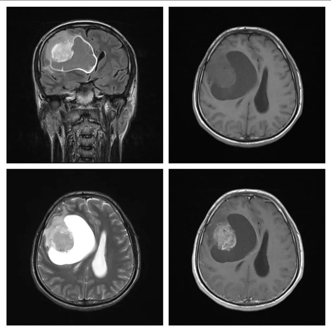
What is the most likely diagnosis on these images of a 21 years old male patient who had presented with headache, vomiting, and left hemiparesis?
- Glioblastoma
- Pilocytic astrocytoma
- Hemangioblastoma
- Lymphoma
- Pleomorphic xanthoastrocytoma
Answer
Glioblastoma it is. It is unusual at a young age, but the contrast enhancement pattern and vasogenic edema around the solid component support this diagnosis.
The mural nodules of pilocytic astrocytoma and hemangioblastoma show more homogenous and avid contrast enhancement, and there are flow voids on the T2-weighted image.
Lymphoma is usually solid, contrast-enhancing, periventricular tumor.
Pleomorphic xanthoastrocytoma can be a grade 2 or 3 tumors. It was a very close differential diagnosis, and difficult to differentiate radiologically from Glioblastoma on these images.