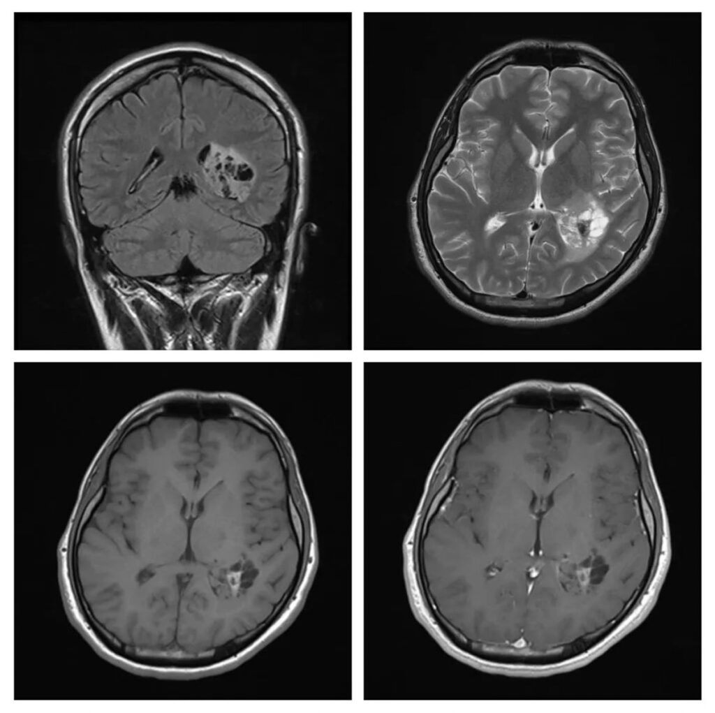
What is the likely diagnosis on these scans of a 25 years old patient who had presented with headaches?
- Trigonal meningioma
- High-grade astrocytoma
- Tanycytic ependymoma
- Lymphoma
- Choroid plexus carcinoma
Answer
Congratulations to those who thought its a tanycytic ependymoma. Ependymoma is often contrasted with enhancing lesions, but lower-grade ependymoma of the tanycytic histological subtype can be non-enhancing. The intraventricular location, cystic cum solid appearance, intra-lesional hemorrhage, and calcification are also features of ependymoma. The biopsy of this tumor was reported as grade II tanycytic ependymoma.
Meningioma, lymphoma, high-grade astrocytoma, and choroid plexus carcinoma all show contrast enhancement. High-grade lesions are also surrounded by edema.