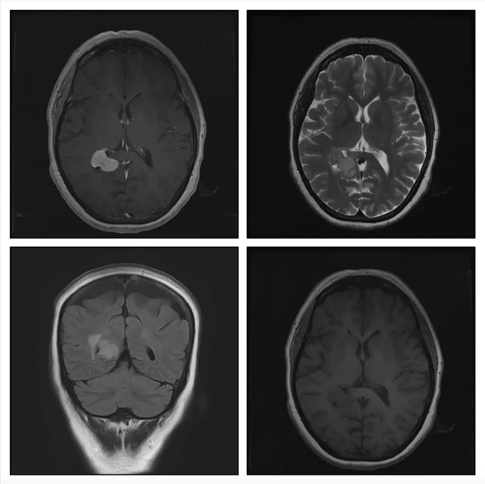
What is the most likely diagnosis in these scans of a 55-year old lady?
- Lymphoma
- Trigonal meningioma
- Glioblastoma
- Ependymoma
- Choroid plexus carcinoma
Answer
Congratulations to all those who guessed trigonal meningioma. These are homogenously enhancing uncommon lesions, and occur in adults. 80% of intraventricular meningioma are present in the trigone of lateral ventricles. They are isointense on T1- and T2-weighted images.
Lymphoma are often homogenously enhancing lesions, present in periventricular area and corpus callosum. They have surrounding edema. There have been few case reports of intraventricular lymphoma.
Glioblastoma are high grade intra-axial tumors that are rarely present within the ventricles. They have heterogenous contrast-enhancement, with central necrotic area, and significant surrounding edema.
Choroid plexus carcinoma are usually present in children and are mostly associated with hydrocephalus. They are avidly contrast enhancing, and often have a frond-like appearance.
Ependymoma is a close differential but they often have calcification and cystic component. They are usually hypointense on T1- and hyperintense on T2-weighted images, and are more common in younger patients.

