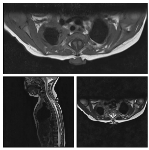
What is the diagnosis in these images?
- Myelomeningocele
- Diastematomyelia & tethered cord
- Syringomyelia
- Intradural extramedullary lesion
- Lipoma of the cord
Answer
Congratulations to all those who selected diastematomyelia and tethered cord. It is a congenital condition in which the spinal cord is split into two halves within a single dural covering. It is often due to a bony spur growing out from the vetebrae, but it can be due to a lesion such as a lipoma, dermoid or epidermoid cyst.
Myelomeningocele would have a CSF filled sac with neural contents. It is most common in lumbosacral region.
There is syringomyelia in these images but that is not the main diagnosis. It is a result of tethering of the cord.