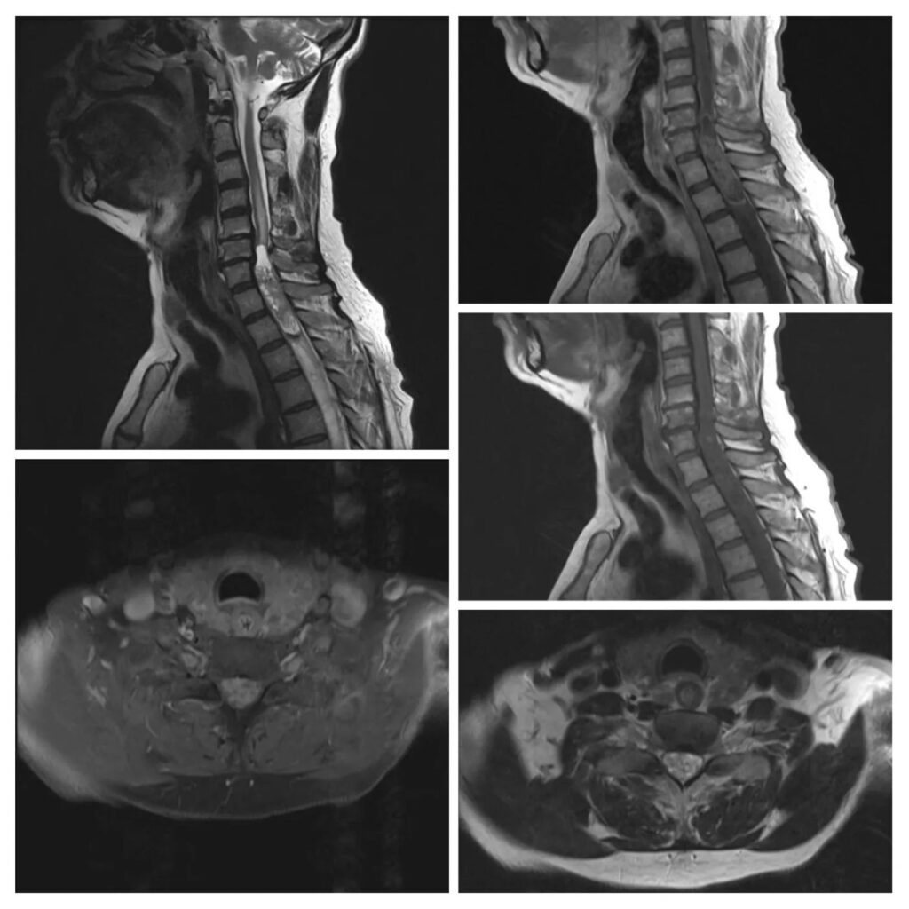
What is the most likely diagnosis in these images of a 63 years old man, with a history of renal transplant?
- Astrocytoma
- Ependymoma
- Metastasis
- Schwannoma
- Hemangioblastoma
Answer
Congratulations to all who selected Ependymoma. It is an intramedullary lesion, which is well-circumscribed. It is iso- to hypo-intense on T1, and hyper-intense on T2, and shows contrast enhancement. There is a hypo-intense cap sign (hemosiderin rim) which signifies associated hemorrhage. The T2-weighted image shows surrounding edema and syringomyelia.
Astrocytoma was a close differential. It is often ill-defined and not well-circumscribed, heterogeneously enhances contrast, and usually does not have associated hemorrhage.
Spinal metastasis is rarely intramedullary and is often dural-based.
Spinal schwannoma is an extra-medullary tumor.
Hemangioblastoma is the 3rd most common intramedullary spinal cord tumor. Larger lesions mostly show flow voids, and there is often a homogenously enhancing nodule.