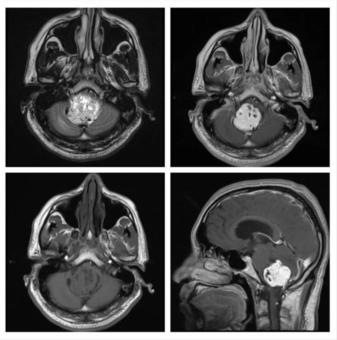
What is the most likely diagnosis in these scans of a 34 years old male patient?
- Medulloblastoma
- Ependymoma
- Metastatic lesion
- Hemangioblastoma
- Dermoid cyst
Answer
Congratulations to all those who guessed Hemangioblastoma. It is mostly cystic cum solid with homogenous contrast enhancement of the solid part, and the most common location is posterior fossa. The solid component is hypo- to iso-intense on T1 and hyperintense on T2 with flow voids due to enlarged vessels. The cystic part is often larger in size with a small mural nodule, but in some cases there are small cysts within the large solid tumor. Cyst walls do not enhance. These are grade 1 lesions, and typically occur in young to middle-aged adults.
Medulloblastoma arises from the roof of 4th ventricle, and occurs mostly in children; only rarely in adults. It is usually solid with heterogenous contrast enhancement and heterogenous signals on T2 because of calcifications and necrosis.
Ependymoma arises from the floor of 4th ventricle, and is a close differential of medulloblastoma. Posterior fossa ependymoma occur mostly in children, and show avid contrast enhancement.
Metastatic lesion usually shows ring enhancement, occurs in older age, and has significant surrounding edema.
Dermoid cyst usually do not enhance on contrast scan and have a predominant cystic component.