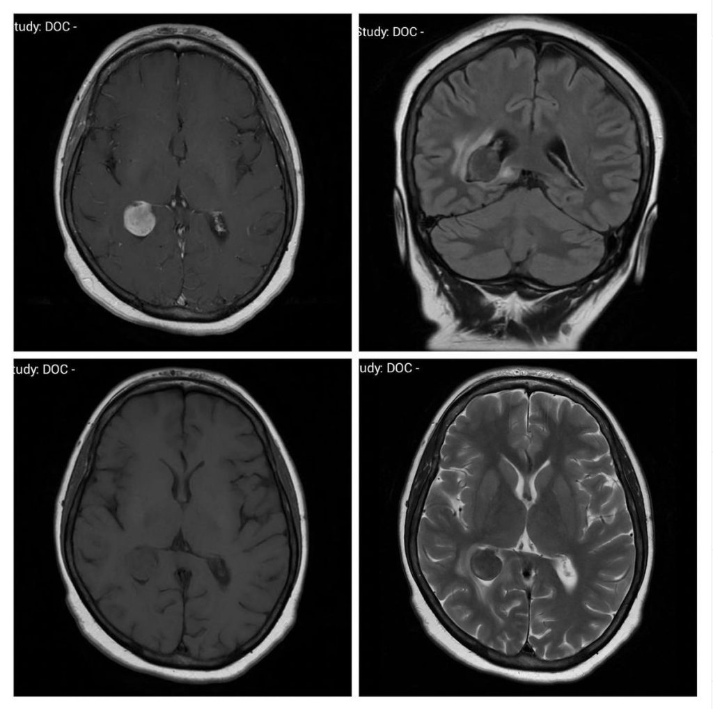
What is the most likely diagnosis on these MRIs of a 56-year-old lady?
- Choroid plexus carcinoma
- Trigone meningioma
- Ependymoma
- Metastasis
- Central neurocytoma
Answer
Congratulations to those who selected trigone meningioma. Intraventricular meningiomas are rare and are most commonly found at the trigone of the lateral ventricle (80%). Their appearance is the same as that of the dural-based meningioma: iso-intense on T1, hyper-intense on T2, and homogenously contrast enhancing, with limited or no cerebral edema.
Choroid plexus carcinoma is iso- to hypo-intense on T1 and T2 and has vivid but heterogeneous contrast enhancement. It occurs more commonly in childhood.
Ependymoma is a close differential with a nearly similar appearance. It is also more common in the younger population and can have cystic areas.
Metastasis is rarely intraventricular. It usually has a central necrotic core and surrounding edema.
Central neurocytoma typically arises from septum pellucidum and shows mild contrast enhancement if any. It is iso-intense on T1 and T2 images.

