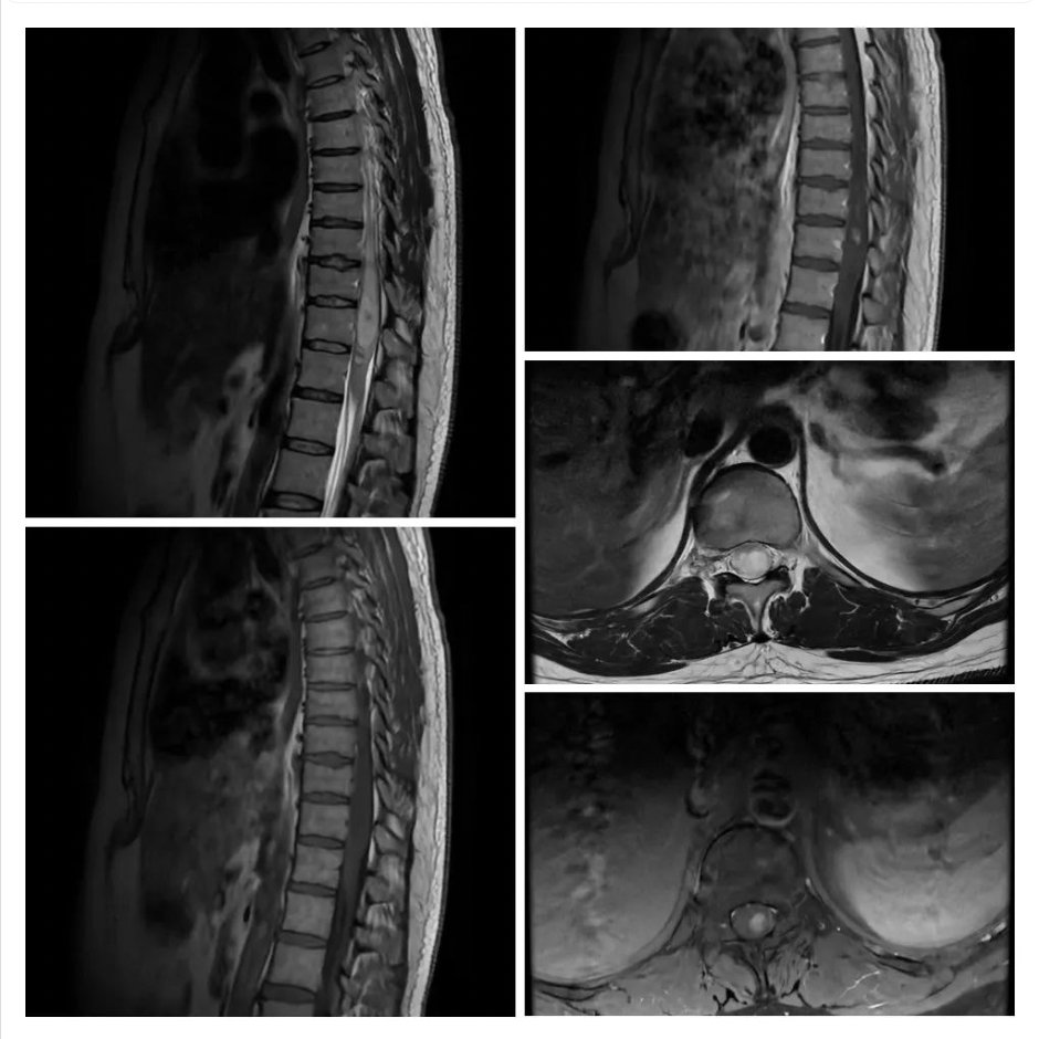
What is the most likely diagnosis in these scans of a 36 years old patient who had presented with back pain and numbness in bilateral lower extremities?
- Astrocytoma
- Ependymoma
- Metastasis
- Spinal Lipoma
- Spinal AVM
Answer
Congratulations to all who chose ependymoma. It is the most common spinal cord tumor. It is intramedullary, often well-circumscribed and central, iso- to hypo-intense on T1 image, hyperintense on T2 image with surrounding edema, and heterogeneously contrast-enhancing. A quarter of spine ependymoma has associated hemorrhage which appears as a hypo-intense hemosiderin rim on the T2 image, called cap sign.
Spinal astrocytoma is the second most common spinal cord tumor and is also intramedullary. It closely mimics ependymoma and is often difficult to differentiate radiologically because of the same signals on MRI. Astrocytoma often has an eccentric location in the spinal cord, whereas ependymoma has a central location.
Metastasis can be intramedullary, but more often they are extramedullary or extradural. There is usually a history of cancer.
Lipoma of the spinal cord appears hyperintense on the T1 and T2 images.
AVM can be intramedullary with surrounding edema but there are hypo-intense flow voids on the T2 image and contrast enhancement.