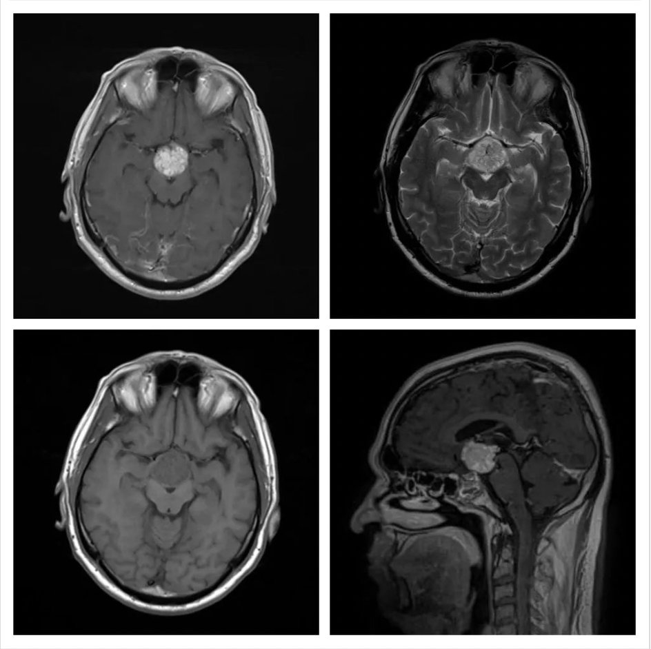
Guess the most likely diagnosis on these images of a 22 years old male patient who had presented with hypopituitarism.
- Planum sphenoidal meningioma
- Pituitary adenoma
- Craniopharyngioma
- Tuberculum sella meningioma
- Suprasellar arachnoid cyst
Answer
Congratulations to those who guessed craniopharyngioma. These are suprasellar lesions, which are more common in children and often cystic. The papillary type is found in adults and is usually solid. It is iso- to slightly hypo-intense on T1, has variable intensity on T2, and has vivid contrast enhancement.
Planum sphenoidal meningioma is iso-intense on T1 and T2, arising from the roof of the sphenoid sinus. It shows avid contrast enhancement and often has a dural tail and CSF sleeve. It pushes the optic nerves posteriorly and can cause visual impairment.
Pituitary adenoma arises from the pituitary gland, causes widening of the sella, and shows heterogenous enhancement due to cystic areas.
Tuberculum sellae meningioma arises in the anterior cranial fossa from the dura of the tuberculum sellae, prechiasmatic sulcus, or diaphragma sellae. Because of its close vicinity to the optic nerves, it causes visual symptoms early.
A suprasellar arachnoid cyst has the same signals as that of CSF with no contrast enhancement.