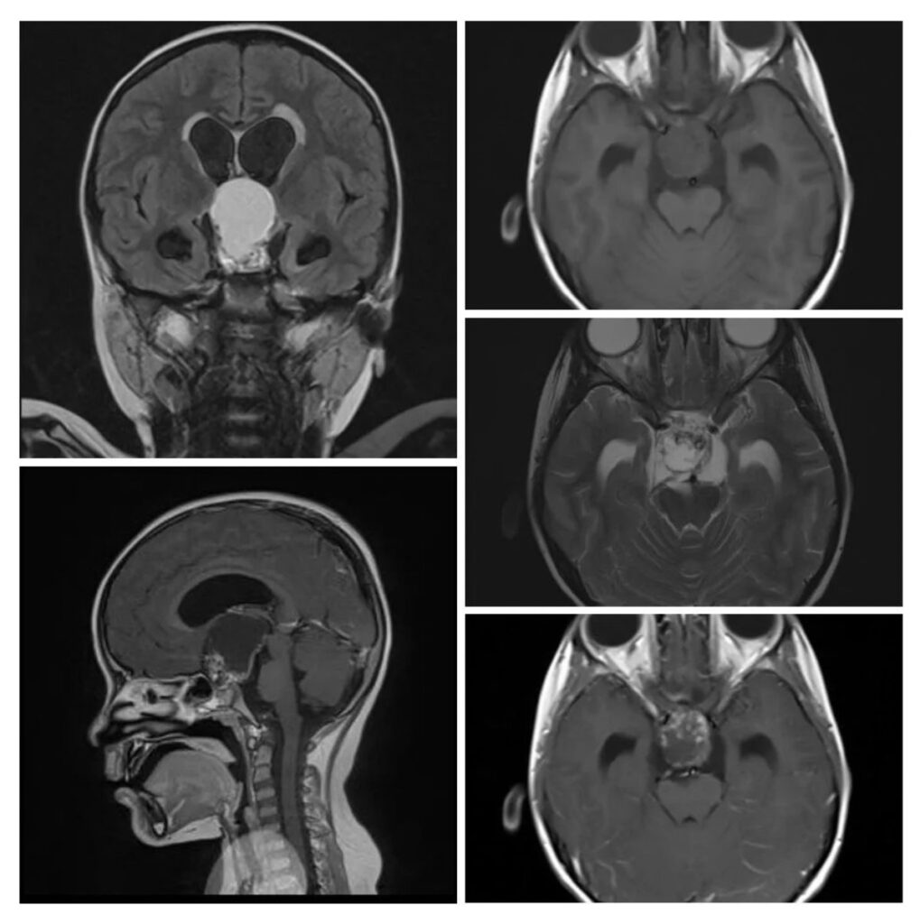
What is the most likely diagnosis on these images of a 10 years old girl who had presented with headache, vomiting, and hypopituitarism?
- Pituitary macroadenoma
- Arachnoid cyst
- Dermoid cyst
- Craniopharyngioma
- Germ cell tumor
Answer
Congratulations to all those who guessed craniopharyngioma. The young age of this patient, cystic cum solid consistency of the lesion, calcification on T2-weighted image, and heterogenous enhancing pattern support this diagnosis in radiology.
Pituitary macroadenoma is often homogenously enhancing, usually solid, causing widening of the sella and occurring in the middle ages.
Arachnoid cyst has the same signals as CSF with no contrast enhancement.
Germ cell tumor is mostly solid, iso-intense on T1- and T2-weighted images, and shows avid contrast enhancement.

