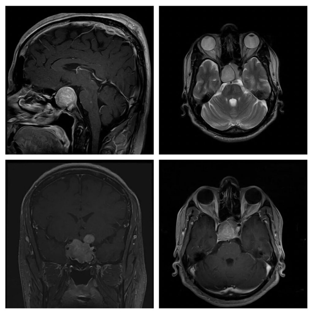
What is the most likely diagnosis on these scans of a 44 years old male patient who had presented with visual impairment and headache?
(Note: The lesion was iso-intense on T1)
- Craniopharyngioma
- Tuberculum sella meningioma
- Pituitary macroadenoma
- Sellar arachnoid cyst
- Planum sphenoidal meningioma
Answer
Congratulations to those who chose pituitary macroadenoma. The enhancement pattern, widening of sella, and extension towards the sphenoid sinus were important findings.
Tuberculum sella meningioma has an almost similar enhancement pattern, but it grows more towards the suprasellar region and there is often a dural tail visible.
Planum sphenoidal meningioma is located in the anterior skull base and extends posteriorly and superiorly. It can extend to sella but that is not the epicenter.
Craniopharyngioma is sellar, suprasellar, or parasellar, but often has heterogenous enhancement with calcifications, and can be cystic cum solid. They are more common at a younger age.
Sellar arachnoid cyst has the same signals as that of CSF and does not show contrast enhancement.