Select the most likely diagnosis from the options below.
- Meningocele and hydrocephalus
- Lipomyelomeningocele and communicating hydrocephalus
- Spina bifida occulta
- Myelomeningocele and aqueductal stenosis causing hydrocephalus
- Myelomeningocele and Chiari malformation
Answer
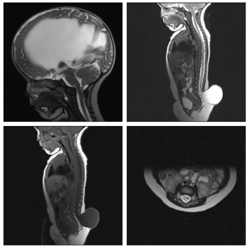
Congratulations to all those who guessed myelomeningocele and aqueductal stenosis causing hydrocephalus (option D). The sagittal section of the T2-weighted image shows tethering of the spinal cord. The neural tissue can be seen going towards the upper border of the sac, at the level of the L5 vertebra. There is associated syringomyelia as well. In the cranial sagittal section of the T2-weighted image, the lateral ventricle and 3rd ventricle are huge (hydrocephalus), while the 4th ventricle is small, and this depicts aqueductal stenosis.
In a meningocele, there is usually no tethering of the spinal cord, and only a CSF-filled sac lined by dura is present.
A lipomyelomeningocele would have the lipoma appear hyperintense on a T1-weighted image.
In communicating hydrocephalus, all ventricles are proportionately enlarged.
Spina bifida occulta involves missing posterior elements of the vertebral column but no herniation of dura.
In Chiari malformation, one or more contents of the posterior fossa herniate below the foramen magnum towards the cervical spinal canal.