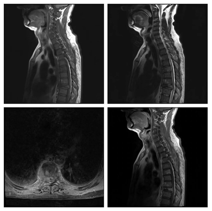
Select the most likely diagnosis for the the radiology images of a 45 years old patient.
- Tuberculosis
- Metastasis
- Vertebral osteomyelitis
- Aneurysmal bone cyst
- Chordoma
Answer
Congratulations to all those who had guessed metastasis. Spine metastasis commonly present with osteolytic compression fractures, show contrast enhancement, and involve both anterior and posterior columns as seen in these images. The discs are usually spared.
Tuberculosis and other infective osteomyelitis involve the endplates of vertebrae, extend into disc space, and often have an epidural or paravertebral component.
Aneurysmal bone cyst does not show homogenous contrast enhancement, appears like a cystic change in the bone, and occasionally its margin shows subtle contrast enhancement.
Chordoma is a locally aggressive tumor which appears as a firm mass with some necrotic areas. It occurs commonly in the sacrococcygeal area and is less frequent in thoracic spine. It extends across the disc and involves more than one vertebra. There is usually an epidural component as well with compression on neural structures.

