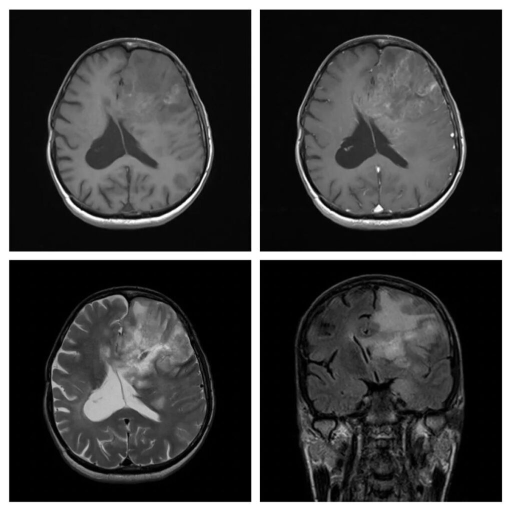
What is the most likely diagnosis on these set of images in a 45 years old patient who had presented with dysphasia and right hemiparesis?
- Low-grade astrocytoma
- Glioblastoma
- Lymphoma
- Ischemic infarct
- Abscess
Answer
Congratulations to all those who selected Glioblastoma. The infiltrative enhancement pattern, extension to the corpus callosum, significant edema, and mass effect are the factors that favor this diagnosis. The hyperintense signals in the T1-weighted image are likely hemorrhage within the lesion, which is a characteristic of a high-grade lesion.
Low-grade astrocytoma usually does not enhance and does not extensive edema. Lymphoma is periventricular and mostly homogenously enhancing lesions. Ischemic infarct will involve a vascular territory. An abscess will show a ring enhancement pattern with central hypointensity.

