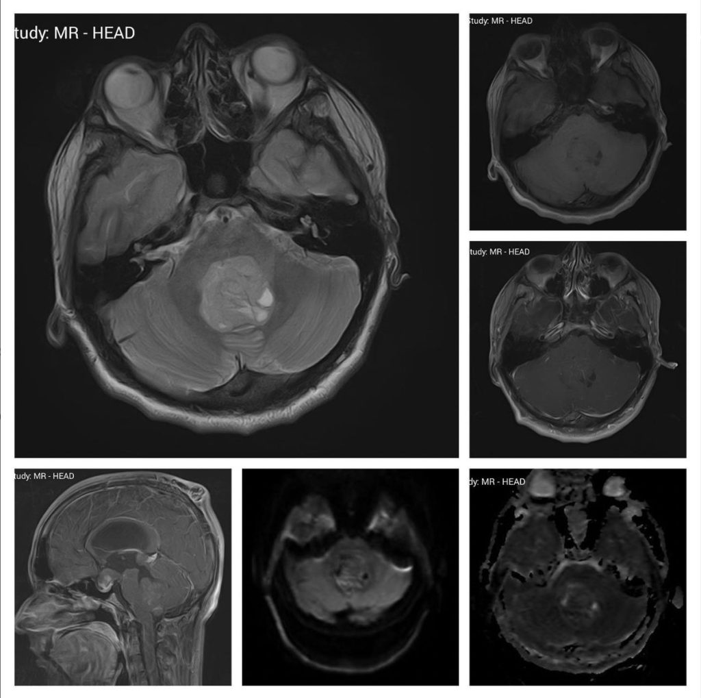
Select the most likely diagnosis on these MRI scans of a 10 years old boy.
- Ependymoma
- Choroid Plexus Carcinoma
- Medulloblastoma
- High-Grade Glioma
- Hemangioblastoma
Answer
Congratulations to those who selected Medulloblastoma. This is the second most common childhood malignant brain tumor. It commonly arises from the roof of the 4th ventricle, is hypo-intense on T1, hyper-intense on T2, and shows heterogenous contrast enhancement. Only 10% of Medulloblastoma does not enhance on post-contrast images. They may or may not show diffusion restriction.
Ependymoma usually arises from the floor of the 4th ventricle, often moving out from the foramen of Luskha, and generally has avid contrast enhancement.
Choroid plexus carcinoma is more often found in the lateral ventricles and causes avid contrast enhancement.
High-grade glioma is uncommon in children. Pilocytic astrocytoma is more common but shows an enhancing nodule with a large cyst.
Hemangioblastoma is usually cystic with an enhancing mural nodule. It can also be solid with homogenous contrast enhancement.

