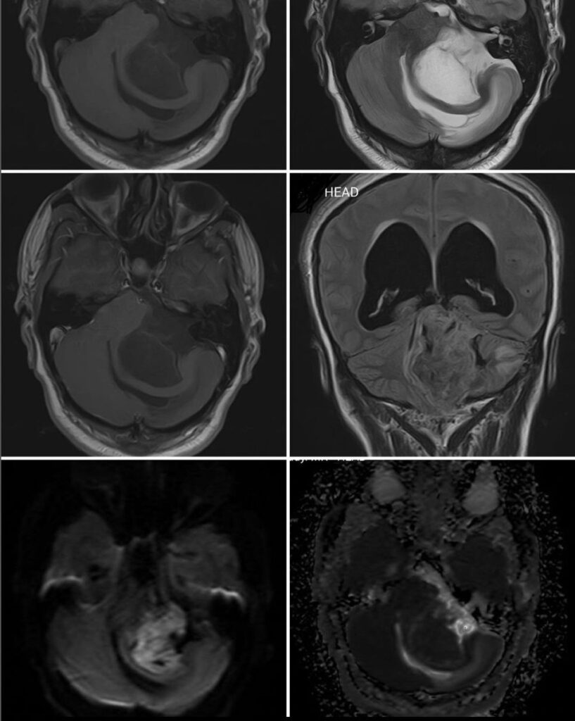
What do you think is the most likely diagnosis on these scans of a 32-year-old patient?
- Acoustic Schwannoma
- Arachnoid cyst
- Meningioma
- Epidermoid cyst
- Hemangioblastoma
Answer
Congratulations! It is an Epidermoid cyst. The contents are like CSF on T2, but there is subtle hyper-intensity on T1 as compared to the CSF in the 4th ventricle. It has diffusion restriction (bright on DWI + dark on ADC). It is heterogenous on FLAIR (not following CSF signals).
An arachnoid cyst has similar signals as CSF.
A cystic acoustic schwannoma shows contrast enhancement in its wall and often has a solid component as well.
A meningioma usually has homogenous contrast enhancement and a dural tail.
Hemangioblastoma is commonly cystic but there is a contrast-enhancing mural nodule. They are usually intra-axial.

